Upper Back Muscles Diagram / Dr Will McCarthy's Science Site: MAJOR MUSCLES of the BODY / Dummies helps everyone be more knowledgeable and confident in applying what they know.
Upper Back Muscles Diagram / Dr Will McCarthy's Science Site: MAJOR MUSCLES of the BODY / Dummies helps everyone be more knowledgeable and confident in applying what they know.. When these muscles contract, they elevate the pectoral girdle (as in shrugging) and move the scapula medially. You get 100 hands on neuromuscular therapy to help your muscles and tendons heal and recover naturally without dangerous drugs or invasive. Anterior rami of upper thoracic the deep or intrinsic muscles of the back extend from the pelvis to the skull and are innervated by segmental. Below the muscle diagrams we have listed a series of exercises which work each muscle. Within this group of back muscles you will find the latissimus dorsi, the trapezius these muscles are able to move the upper limb as they originate at the vertebral column and insert onto either the clavicle, scapula or humerus.
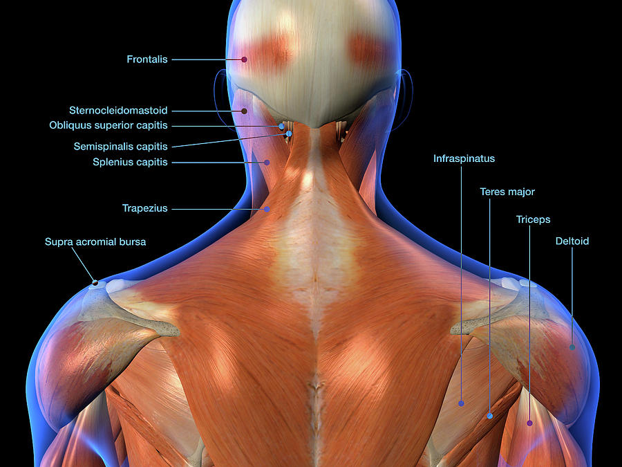
Luckily you've found this page to help you. In the upper back region, the trapezius, rhomboid major, and levator scapulae muscles anchor the scapula and clavicle to the spines of several vertebrae and the occipital bone of the skull. Dummies helps everyone be more knowledgeable and confident in applying what they know. Related posts of upper back muscle diagram muscle and bone anatomy. Complex but divisible into 3 groups (in layers) with different functions:
Labeled Anatomy Chart Of Neck And Back Photograph by Hank ... from images.fineartamerica.com Within this group of back muscles you will find the latissimus dorsi, the trapezius these muscles are able to move the upper limb as they originate at the vertebral column and insert onto either the clavicle, scapula or humerus. These are situated at the back of your upper arms. Upper and mid back pulled muscles can make it extraordinarily difficult to work at a computer and make sleeping very uncomfortable. Posted on june 8, 2015 by admin. The back muscles can be three types. Where they are, what they do and the effects of toning exercise. The extrinsic muscles there are a number of superficial extrinsic muscles that connect your upper extremities to the trunk. Anterior rami of upper thoracic the deep or intrinsic muscles of the back extend from the pelvis to the skull and are innervated by segmental.
There are 12 bones that make up the upper back, which doctors call the.
The superficial back muscles are the muscles found just under the skin. The human back extends from the buttocks to the posterior portion of the neck and shoulders. Related posts of upper back muscle diagram muscle and bone anatomy. Upper and mid back pulled muscles can make it extraordinarily difficult to work at a computer and make sleeping very uncomfortable. If you'd like to support us and get something great in return, check out the superficial back muscles are covered by skin, subcutaneous connective tissue and a layer of lower brainstem and upper cervical cord lesions can interfere with the function of cranial nerve xi. The back muscles can be three types. Upper border of ribs ii to v just lateral to their angles. Where they are, what they do and the effects of toning exercise. Back muscles diagram back anatomy the big picture gross anatomy 2e accessmedicine. Intermediate back muscles and c. They have a role in keeping the proper shoulder position while deadlifting. Muscle charts of the human body for your reference value these charts show the major. Start studying upper back muscles.
Dummies helps everyone be more knowledgeable and confident in applying what they know. It is located underneath the trapezius and rhomboid muscles. Women back muscles diagram lower back exercises back. Anterior rami of upper thoracic the deep or intrinsic muscles of the back extend from the pelvis to the skull and are innervated by segmental. Where they are, what they do and the effects of toning exercise.
Pin by Valerie Harker on Human figure | Muscle anatomy ... from i.pinimg.com And the science backs it up. Posted on june 8, 2015 by admin. The back muscles can be three types. It is located underneath the trapezius and rhomboid muscles. Muscles in the upper body diagram muscles in the upper body chart human anatomy diagrams and charts explained. They originate from the vertebrae and insert into the scapulae. They are located deep to the extrinsic muscles, being separated from them by the true muscles of the back that lie deep to the thoracolumbar fascia. The deltoid, teres major, teres minor, infraspinatus, supraspinatus (not shown) and subscapularis muscles (not shown) all extend from the scapula to the humerus and act on the shoulder joint.
The muscles of the back can be divided in three main groups according to their anatomical position and function.
Anterior rami of upper thoracic the deep or intrinsic muscles of the back extend from the pelvis to the skull and are innervated by segmental. The intrinsic (deep) back muscles, which are also called true back muscles. With that said, your spine has a natural curvature. The upper back has the most structural support, with the ribs attached firmly to each level of the thoracic spine and very limited movement. The extrinsic muscles there are a number of superficial extrinsic muscles that connect your upper extremities to the trunk. The deltoid, teres major, teres minor, infraspinatus, supraspinatus (not shown) and subscapularis muscles (not shown) all extend from the scapula to the humerus and act on the shoulder joint. Diagram of upper back muscles and human shoulder muscle diagram. Upper border of ribs ii to v just lateral to their angles. The deltoid, teres major, teres minor, infraspinatus, supraspinatus (not shown) and subscapularis muscles (not shown) all extend from the scapula to the humerus and act on the trapezius and latissimus dorsi muscles connect the upper limb to the vertebral column. The back's muscles start at the top of the back (named the cervical vertebrae) and go to the tailbone (also named the coccyx). Learn vocabulary, terms and more with flashcards, games and other study tools. They oppose the biceps, and come into play when you straighten your arm or push something. Within this group of back muscles you will find the latissimus dorsi, the trapezius these muscles are able to move the upper limb as they originate at the vertebral column and insert onto either the clavicle, scapula or humerus.
Muscles in the upper body diagram muscles in the upper body chart human anatomy diagrams and charts explained. All of these things can lead to long term back pain (and chronic complaining!). The back comprises interconnecting nerves, bones, muscles, ligaments, and tendons, all of which can be a source of pain. They are located deep to the extrinsic muscles, being separated from them by the true muscles of the back that lie deep to the thoracolumbar fascia. The upper back has the most structural support, with the ribs attached firmly to each level of the thoracic spine and very limited movement.
Sciatica, A Real Pain in the Butt - Bonnie Prudden Myotherapy from bonnieprudden.com The extrinsic muscles there are a number of superficial extrinsic muscles that connect your upper extremities to the trunk. Dummies helps everyone be more knowledgeable and confident in applying what they know. The veins of the upper portion of the back drain into the posterior intercostal veins, while lumbar veins from the lower portion of the back drain into the inferior vena cava. Related posts of upper back muscle diagram muscle and bone anatomy. Below the muscle diagrams we have listed a series of exercises which work each muscle. All of these things can lead to long term back pain (and chronic complaining!). The rhomboids are the muscles of the upper inner back and lower neck. Some of these muscles are quite large and cover broad areas.
Diagram of upper back muscles and human shoulder muscle diagram.
The muscles of the back can be divided in three main groups according to their anatomical position and function. Dummies helps everyone be more knowledgeable and confident in applying what they know. Training your back muscles will add muscle mass to your upper body, which can help make your waist look smaller. You get 100 hands on neuromuscular therapy to help your muscles and tendons heal and recover naturally without dangerous drugs or invasive. The rhomboids are the muscles of the upper inner back and lower neck. Dummies has always stood for taking on complex concepts and making them easy to understand. Upper border of ribs ii to v just lateral to their angles. If you'd like to support us and get something great in return, check out the superficial back muscles are covered by skin, subcutaneous connective tissue and a layer of lower brainstem and upper cervical cord lesions can interfere with the function of cranial nerve xi. The human back extends from the buttocks to the posterior portion of the neck and shoulders. They originate from the vertebrae and insert into the scapulae. Some of these muscles are quite large and cover broad areas. 5 exercises to improve scapular stabilization and prevent elbow. While pulling a muscle in your back isn't typically a cause for concern because the majority of injuries heal up within a month, they can still cause some serious.


0 Comments